The Turning Point: Real-World Stories Where an ECG Changed Everything – Case 1: Pericardial Effusion
Welcome to our new series, “The Turning Point,” where we spotlight real-world cases from your colleagues that demonstrate how an ECG can dramatically alter a patient’s outcome. Today’s case is a powerful reminder of why a pre-anesthetic ECG is more than just a checkbox.
The Patient: A middle-aged Golden Retriever scheduled for surgery for IVDD (Intervertebral Disc Disease).
The Presenting Signs: The dog was otherwise stable, with the neurological deficits being the primary focus. The routine physical exam was unremarkable.
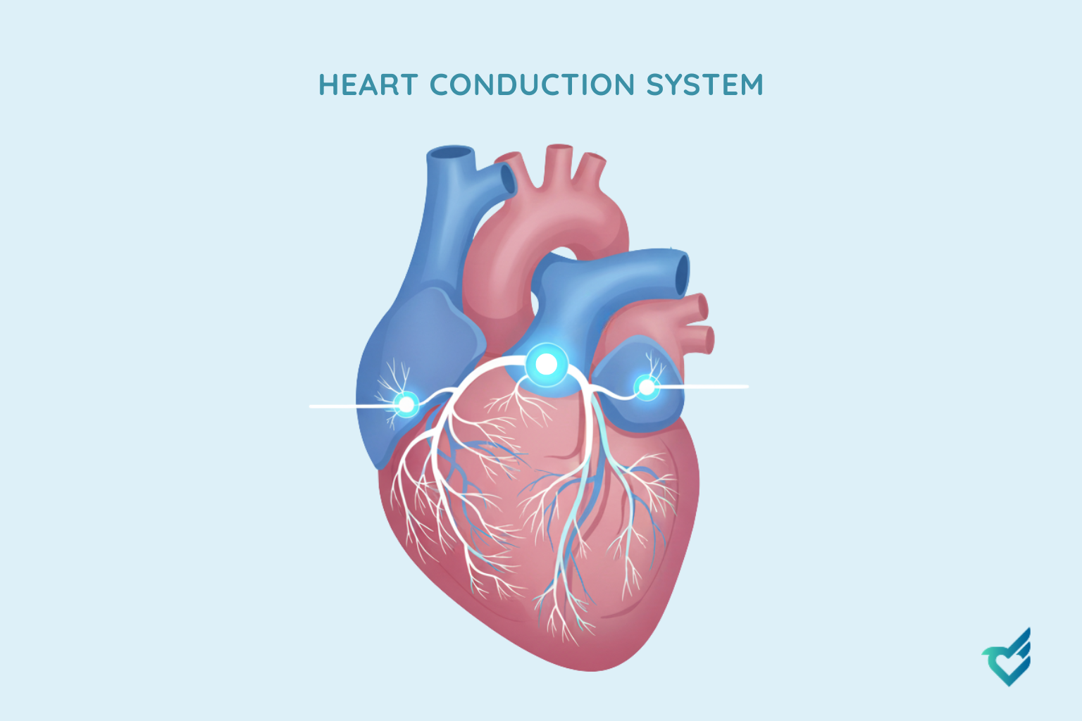
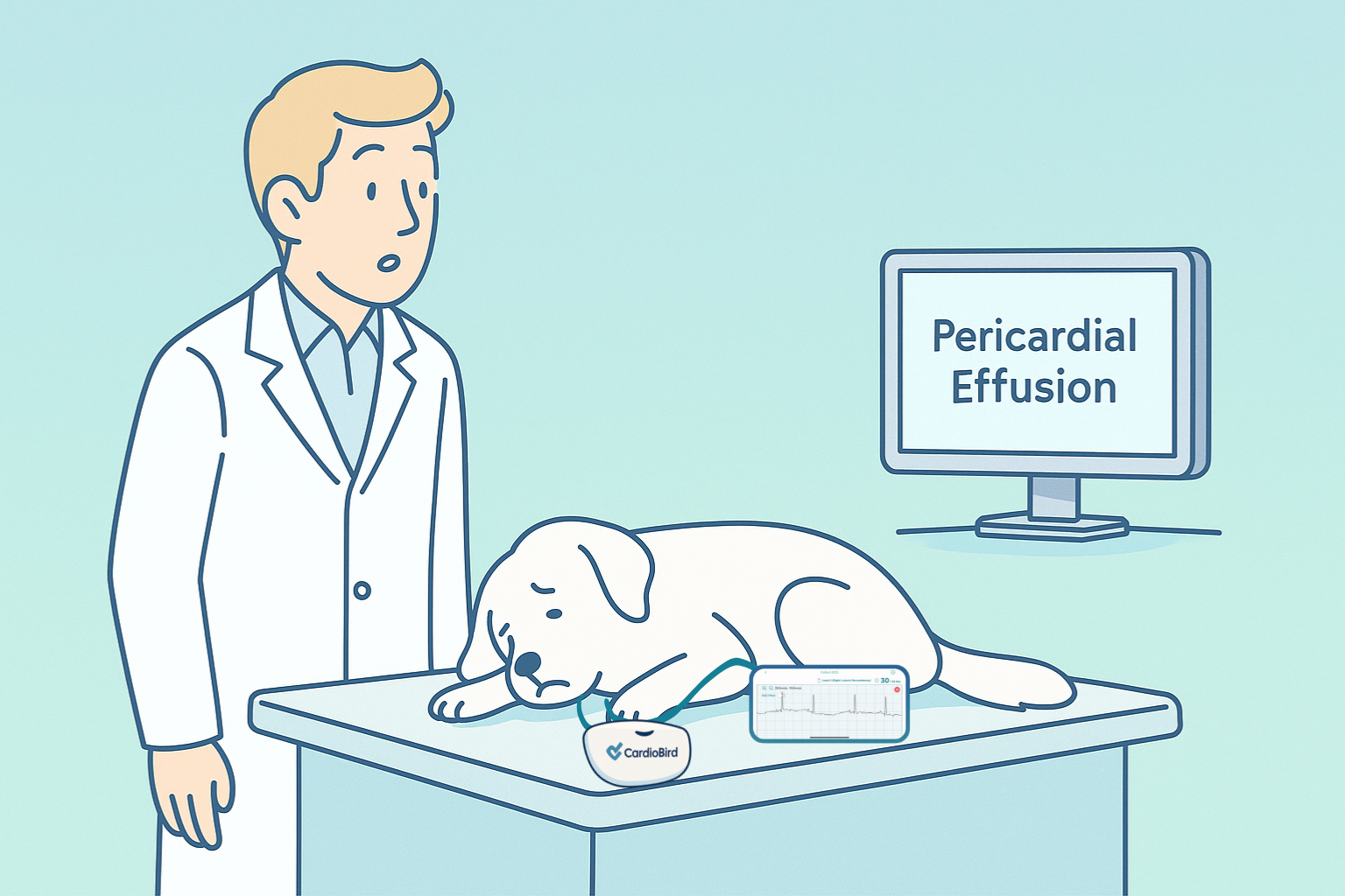
The Turning Point – The Pre-Anesthetic ECG: The attending veterinarian, a CardioBird user, performed a pre-anesthetic ECG. The initial trace from a single lead was concerning, showing subtle signs suggestive of pericardial effusion: low-voltage QRS complexes and what appeared to be electrical alternans.
ECG Findings & Critical Difference:
A single-lead ECG cannot definitively confirm pericardial effusion, but it provides the crucial clues to warrant immediate action. In this case, the veterinarian followed a key protocol: they ran repeated tracings, specifically on both lead I and lead III. The consistent presence of low-voltage QRS complexes (<1.0 mV) across these leads, combined with the characteristic “swinging” heart pattern of electrical alternans visible in multiple leads, provided strong, multi-lead evidence supporting the suspicion.
This ECG evidence was the first and only clue. Without it, the dog would have proceeded to anesthesia, where the cardiovascular effects of drugs could have precipitated a catastrophic crisis. Guided by the compelling multi-lead ECG findings, the team performed an echocardogram, which confirmed a significant pericardial effusion. The surgical plan was halted, and the patient’s life was saved.
Your Takeaway:
This case underscores critical lessons for primary care practice:
- The ECG is a Screening Tool: It raises suspicion for pericardial effusion but is not a standalone diagnostic. If you see low-voltage complexes or electrical alternans in one lead, don’t stop there.
- Run Multiple Leads: For a more confident assessment, run tracings on both lead I and lead III. Consistent findings across multiple leads significantly increase the index of suspicion.
- Guide Your Next Steps: This ECG pattern is your signal to postpone elective anesthesia and pursue definitive diagnosis via echocardiography, even if it means a referral. It directly alters case management and mitigates risk.
You are part of a global community using AI-driven technology to practice smarter, safer medicine. Together, we are ensuring that no critical clue goes unnoticed.
Have a “Turning Point” case of your own? We’d love to feature it. Share your story with us.