The Influence of Lead placement and Body Position on ECG in Dogs and Cats: A Practical Guide for Veterinarians
Estimated reading time: 3.57 minutes

Welcome back to the CardioBird bi-weekly newsletter! This issue, we’re diving into a topic that’s fundamental to ECG interpretation: the influence of lead placement and body position. While ECGs are invaluable diagnostic tools, understanding how these factors affect the readings is crucial for accurate diagnosis and treatment.
Why Does Lead Placement Matter?
Think of an ECG as capturing electrical activity from different angles. Standard limb leads (I, II, and III) provide distinct perspectives on the heart’s electrical axis.
- Lead I: Records the potential difference between the left and right forelimbs.
- Lead II: Records the potential difference between the right forelimb and the left hindlimb. This lead often provides the clearest P waves and QRS complexes, making it a common choice for initial rhythm assessment.
- Lead III: Records the potential difference between the left forelimb and the left hindlimb.
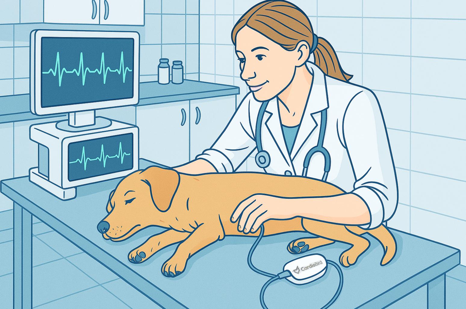
So, what happens when you don’t use standard limb leads? Some wearable ECG devices for pets use alternative placements on the chest or trunk. These placements can still provide valuable data, but it’s important to remember that the waveforms and amplitudes may differ significantly from standard limb lead recordings. This is because the electrical signals are being picked up from a different location relative to the heart. Therefore, when using such devices, make sure to use the reference ranges and interpretation guidance specific to that device, as established diagnostic benchmarks (Tilley, 1992) derived from standard Lead II recordings in the right lateral recumbency position will not be applicable.
The Effect of Body Position on ECG
Body position significantly influences ECG waveforms. Factors such as gravity, electrode contact, and the heart’s orientation within the thoracic cavity can all impact the morphology and amplitude of the ECG.
Common Body Positions in Veterinary ECG:
- Right Lateral Recumbency:
- Gold Standard Position: Provides the most consistent waveforms and is preferred for diagnostic ECGs.
- Why It Works: The heart’s position relative to the electrodes is stable, and artifacts are minimized.
- Clinical Applications: Use for routine diagnostics and when precision is critical (e.g., arrhythmia evaluation).
- Sternal Recumbency:
- Practical for Cats and Small Dogs: Cats, in particular, tolerate this position better than lateral recumbency.
- Effects: May slightly alter wave morphology, such as reducing R-wave amplitude, but remains clinically acceptable.
- Clinical Applications: A good alternative for animals that resist lateral recumbency.
- Sitting Position:
- Useful for Large or Anxious Dogs: Many large dogs find sitting more comfortable than lying down.
- Effects: Studies (e.g., Coleman and Robson, 2005) show greater variability in waveforms, particularly the P wave and QRS complex.
- Clinical Applications: Best for quick rhythm checks or when lateral recumbency is not feasible.
- Standing Position:
- Emergency or Critical Scenarios: May be necessary for patients with respiratory distress or severe anxiety.
- Effects: Introduces the most variability in waveforms and may increase artifacts.
-
- Clinical Applications: Use only when absolutely necessary, prioritizing rhythm evaluation over precise waveform measurements.
Practical Tips for Lead and Body Position Selection
General Guidelines
- Standardization is Key:
- Consistency in lead and body position is critical for reliable interpretations, especially for serial ECGs.
- If using a wearable ECG device, ensure the same placement is used across all recordings.
- Minimize Artifacts:
- Ensure good electrode contact with alcohol or conductive gel.
- Place the patient on a non-conductive surface (e.g., rubber mat).
Choosing the Right Lead and Body Position for Clinical Scenarios
| Clinical Scenario | Recommended Lead and Body Position |
| Routine rhythm evaluation | Lead II, right lateral recumbency (gold standard). |
| Fractious cats or small dogs | Lead II, sternal recumbency. |
| Large or anxious dogs | Lead II, sitting position |
| Emergency cases (e.g., respiratory distress) | Lead II, standing or sternal recumbency. |
| Wearable ECG (non-standard positions) | Ensure consistent placement (e.g., chest/trunk). Focus on rhythm trends rather than precise morphology. |
Final Thoughts
Understanding the effects of lead and body positions on ECG recordings is essential for accurate interpretation. While Lead II in right lateral recumbency remains the gold standard, adapting to patient needs—such as using sternal or sitting positions—ensures you can obtain reliable data in any situation. Wearable ECG devices, with their unique electrode placements, offer exciting opportunities for continuous monitoring, but consistency in placement is key to maintaining accuracy.
At CardioBird, we’re here to help you make the most of your ECG recordings, no matter the clinical scenario.
Stay tuned for more insights in our newsletter!
References:
- Tilley. (1992), Essentials of Canine and Feline Electrocardiography: Interpretation and Treatment
- Rishniw, Mark, et al. “Effect of body position on the 6‐lead ECG of dogs.” Journal of Veterinary Internal Medicine (2002).
- Stern, Joshua A., et al. “Effect of body position on electrocardiographic recordings in dogs.” Australian Veterinary Journal (2013).
- Coleman, M. G., & Robson, M. C. (2005). American Journal of Veterinary Research.
- Sousa et al. (2018). Influence of body position on the measurement of electrocardiographic waves in healthy dogs.
- Harvey et al. (2005). Effect of Body Position on Feline Electrocardiographic Recordings.

