The ECG Decoded: A Veterinarian’s Guide to the Heart’s Rhythm – Part 8: Linking Electrical disturbance to Patient Outcomes: Hemodynamic consequence and related clinical signs
Estimated reading time: 3.48 minutes
Welcome to the final installment of our series, The ECG Decoded: A Veterinarian’s Guide to the Heart’s Rhythm. We have explored the genesis, patterns, and causes of arrhythmias. Now, we complete the circle by answering the most critical question: What does this arrhythmia mean for my patient? This article links electrical disturbances to their hemodynamic consequences and the clinical signs you observe, transforming an ECG finding into an actionable treatment plan.
The Hemodynamic Equation: More Than Just Heart Rate
The impact of an arrhythmia depends on how it disrupts the heart’s primary functions: filling (preload) and ejecting (cardiac output). Three key factors determine the severity:
- Heart Rate: The most obvious factor. Rates that are too slow (< 40-50 bpm in dogs) or too fast (> 240-260 bpm) compromise the time available for filling or ejecting, respectively.
- Rhythm Regularity: The heart relies on predictable intervals. Irregular rhythms like atrial fibrillation or frequent Premature Ventricular Contractions (PVCs) destroy the synchronized atrial “kick” that contributes 15-30% of ventricular filling, reducing stroke volume.
- Coordination of Contraction: This is the most critical factor. In a normal beat, atria contract first to prime the ventricles. In arrhythmias like ventricular tachycardia or complete heart block, this coordination is lost. The ventricles may contract when they are only partially filled, drastically cutting the volume of blood pumped with each beat.
Decoding the Clinical Signs: Why Pets Show Symptoms
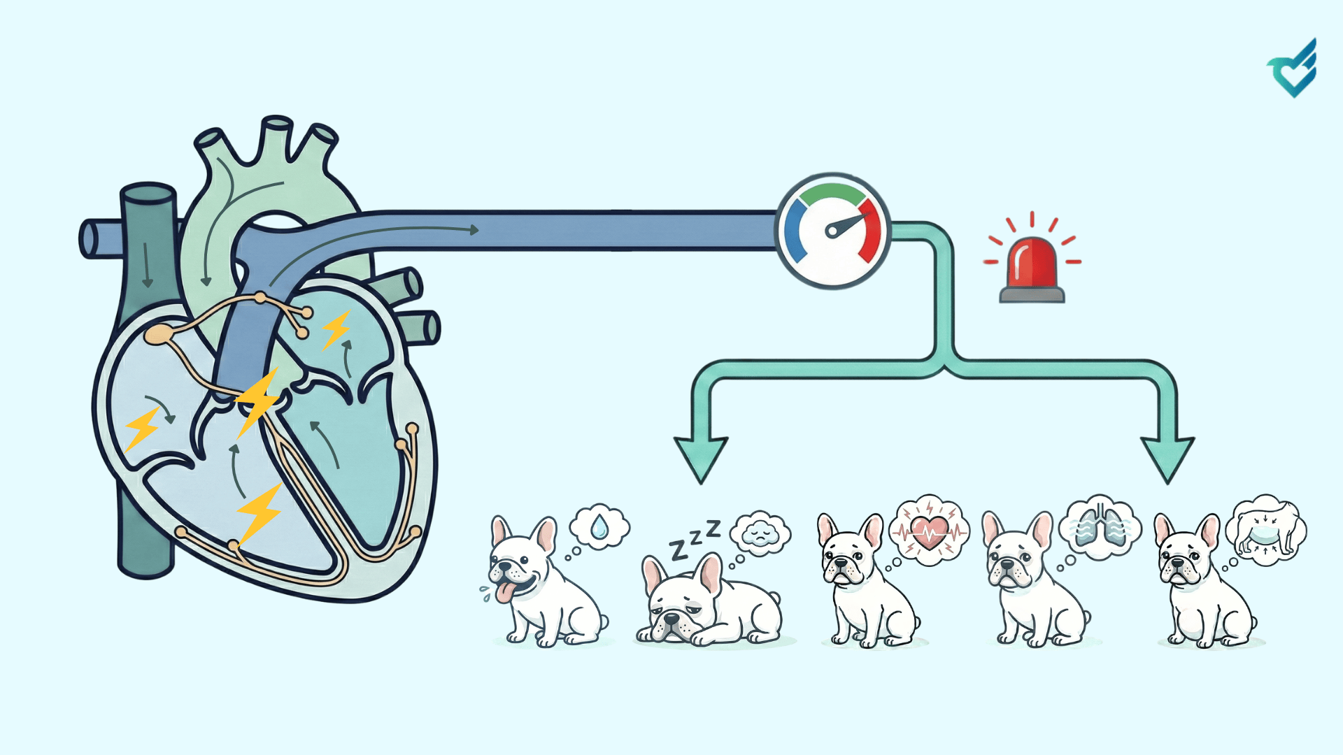
Clinical signs arise directly from reduced cardiac output and poor organ perfusion, or from the neurohormonal activation that tries to compensate for it.
- Signs of Low Cardiac Output & Hypoperfusion:
-
- Syncope (Fainting) & Weakness: Sudden, profound drops in output, as seen with prolonged sinus arrest, ventricular tachycardia, or a very fast Supraventricular Tachycardia (SVT), prevent adequate blood flow to the brain.
- Exercise Intolerance & Lethargy: The heart cannot increase output to meet the demands of activity. This is common with persistent tachyarrhythmias or bradyarrhythmias.
- Pale Mucous Membranes & Prolonged Capillary Refill Time (CRT): Direct signs of poor peripheral perfusion.
- Signs of Compensatory Mechanisms & Congestion:
-
- Tachycardia: The body’s first attempt to maintain output by increasing rate, often seen in response to underlying failure.
- Tachypnea/Dyspnea: Caused by pulmonary edema secondary to left-sided heart failure, which can be triggered or worsened by an arrhythmia like atrial fibrillation.
- Ascites: Fluid buildup in the abdomen from right-sided heart failure, potentially exacerbated by an arrhythmia.
A Practical Framework for Assessing Urgency
Not every arrhythmia requires emergency intervention. Use this combined ECG-clinical assessment to triage:
- Unstable / Emergency: Arrhythmia + Clinical Signs (Syncope, collapse, profound weakness, respiratory distress).
- Examples: Sustained Ventricular Tachycardia, rapid Atrial Fibrillation with signs, complete (3rd-degree) AV block with weakness.
- Action: Immediate therapy (oxygen, antiarrhythmics, electrical cardioversion, pacing) is required to restore perfusion.
- Potentially Unstable / Urgent: Significant arrhythmia + No current signs, but high risk of deterioration.
- Examples: Very fast SVT, frequent multiform PVCs, new-onset atrial fibrillation.
- Action: Requires prompt investigation and treatment to prevent an emergency, but allows time for diagnostics (e.g., echo, electrolytes).
- Stable / Monitor: Arrhythmia + No signs and minimal hemodynamic impact.
- Examples: Occasional uniform PVCs, mild sinus bradycardia in a resting athlete, asymptomatic 1st-degree AV block.
- Action: Investigate underlying cause, monitor, but may not require direct antiarrhythmic therapy.
The CardioBird Advantage: Integrating Signal with Symptom
At CardioBird, our mission has always been to elevate your practice by translating complex data into clinical wisdom. This series has equipped you with the knowledge to decode the ECG. Our AI-ECG service is designed to be your partner in this final, critical step. We don’t just deliver a list of findings; our reports are structured to highlight the arrhythmias with the greatest potential hemodynamic impact, helping you quickly connect the electrical disturbance to your patient’s clinical picture. We empower you to move from detection to decisive action, ensuring you provide care that is not only technologically advanced but profoundly clinically relevant.
Thank you for joining us on this journey through the heart’s rhythm. We are passionate about continuing to provide you with the cutting-edge tools and knowledge that make exceptional care possible.
The CardioBird Team