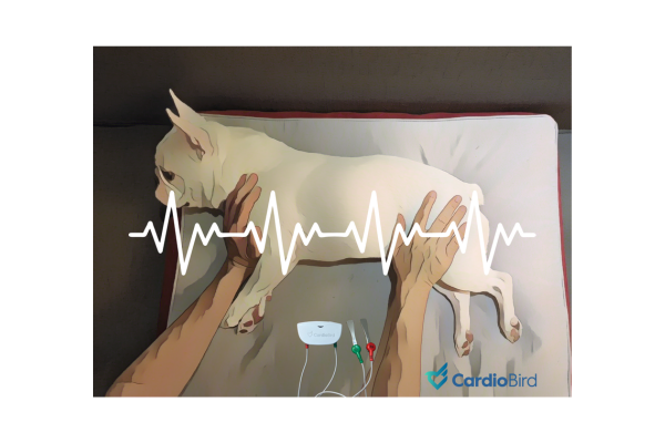
Is Single-Lead ECG Enough for Primary Care Veterinary Practice?

Electrocardiography (ECG) is an essential tool in veterinary medicine, particularly for identifying cardiac rhythm disturbances.
For primary care veterinarians, a single-lead ECG, specifically Lead II, is often sufficient for the rapid diagnosis of most arrhythmias. While multi-lead ECGs provide a more comprehensive assessment of cardiac function, a Lead II ECG offers a practical, efficient, and reliable option in many clinical scenarios. Understanding the strengths and limitations of a Lead II ECG can help veterinarians make informed decisions about its implementation in daily practice.
Why Lead II ECG Is Often Enough
A Lead II ECG records electrical activity along a diagonal axis from the right forelimb to the left hindlimb. This lead is particularly well-suited for evaluating rhythm disturbances because it provides clear visualization of the P wave, QRS complex, and T wave, which are critical components for assessing heart function.
1. Rapid Detection of Arrhythmias
Lead II is highly reliable for identifying common and clinically significant arrhythmias, such as:
- Atrial fibrillation
-
Ventricular tachycardia
-
Premature ventricular and supraventricular contractions
2. Ease of Use
-
Requires minimal setup and is faster to perform compared to multi-lead ECGs.
-
Suitable for use in conscious or lightly sedated patients, making it practical in primary care settings.
3. Cost-Effectiveness
-
Single-lead ECG systems are more affordable and accessible than multi-lead setups, reducing the financial burden on clinics.
4. Versatility in Various Scenarios
-
Ideal for routine health-check and pre-anesthesia screening, intra- and post-anesthesia monitoring, and rapid rhythm evaluation for suspected heart diseases and emergency situations
Limitations of Lead II ECG
While Lead II ECG provides significant diagnostic value for rhythm disturbances, and a high specificity for structural changes detection while measured under right lateral recumbency, it has some limitations. It offers a single-axis perspective and lacks the ability to evaluate the heart’s electrical activity from multiple angles. It is less sensitive than echocardiography for the detection of structural abnormalities.
Lead II ECG can suggest but not definitively diagnose:
- Chamber Enlargement Patterns:
For example, left atrial or right ventricular enlargement may require evaluation from additional leads and/or echocardiography. - Subtle Conduction Abnormalities:
Conditions such as left or right bundle branch block or fascicular blocks are better identified with multi-lead ECGs. - Electrical Axis Deviations:
Despite the ability to detect some form of axis deviation in standard lead II ECG, multi-lead ECG is required when a comprehensive evaluation of electrical axis deviations is indicated.
For these reasons, multi-lead ECGs (such as 6-lead or 12-lead systems) are recommended for cases where more detailed cardiac evaluation is indicated from findings in single-lead ECG or other examinations.
Supporting literature for Single Lead II ECG Use in Primary Care
The use of Lead II ECG in primary care settings is well-supported by the veterinary literature:
- Tilley, L. P. (1992). 《Essentials of Canine and Feline Electrocardiography: Interpretation and Treatment》:
Emphasize that Lead II ECG is the most commonly used configuration for arrhythmia detection in routine practice due to its simplicity and reliability. - Ware (2011). Cardiovascular Disease in Small Animal Medicine: Medicine:
Highlights that Lead II is sufficient for identifying most rhythm disturbances, though multi-lead ECGs are recommended for definitive diagnosis of structural abnormalities and complex conduction issues. - Bonagura & Twedt (2013). 《Kirk’s Current Veterinary Therapy XV》:
Reinforce the practicality of single-lead ECGs for rhythm evaluation in primary care, particularly during anesthesia or emergencies. - Tidholm et al. (2001). Journal of Veterinary Internal:
Demonstrated that while multi-lead ECGs are superior for diagnosing complex conditions, single-lead ECGs effectively capture the majority of clinically relevant arrhythmias.
Practical Recommendations for Primary Care Veterinarians
For most primary care practices, a Lead II ECG provides a reliable and efficient method for diagnosing the majority of cardiac rhythm disturbances. Its ease of use, affordability, and rapid results, especially when powered with recent advancements in AI interpretation, make it an indispensable tool for routine and emergency evaluations.
However, veterinarians should recognize when a more comprehensive assessment is needed. For patients with suspected structural heart disease, unexplained syncope, or complex arrhythmias, referral to a cardiologist or the use of a multi-lead ECG may be warranted. Incorporating Lead II ECG into daily practice allows primary care veterinarians to provide timely and effective cardiac care while reserving advanced diagnostics for more complex cases.
Conclusion
In primary care veterinary practice, a single Lead II ECG is often sufficient for the rapid and accurate diagnosis of most cardiac rhythm disturbances. Its simplicity, cost-effectiveness, especially when paired with reliable AI interpretation make it a valuable diagnostic tool for routine and emergency use. While multi-lead ECGs have their place in detailed and further cardiac evaluations, the Lead II ECG remains the cornerstone of arrhythmia detection in primary care. By understanding its strengths and limitations, veterinarians can confidently integrate this tool into their practice to enhance patient care.


