Special Topic: ECG of the Month – The Asymptomatic Cat with Significant ECG Findings
Estimated reading time: 4.55 minutes
A routine health-check ECG in a clinically normal cat can quietly reveal conduction and structural concerns that would otherwise be missed, and this Maine Coon illustrates why ECG should sit alongside auscultation as a standard screening tool in everyday practice.
Case information
A 4‑year‑old neutered male Maine Coon cat, body weight 5.90 kg, underwent a routine health check with no reported clinical signs. Heart auscultation raised uncertainty about the presence of a murmur, and no obvious arrhythmia was noted on the stethoscope. A standard Lead II ECG was recorded in right lateral recumbency using the CardioBird AI‑ECG as part of the clinic’s wellness protocol.
CardioBird analysis identified a mean heart rate of 131 bpm (range 102–156 bpm), which is slower than the normal reference range of 140–240 bpm for cats, and was classified as sinus bradycardia. The rhythm was irregular, with inconsistent RR intervals and no regular repeating pattern.
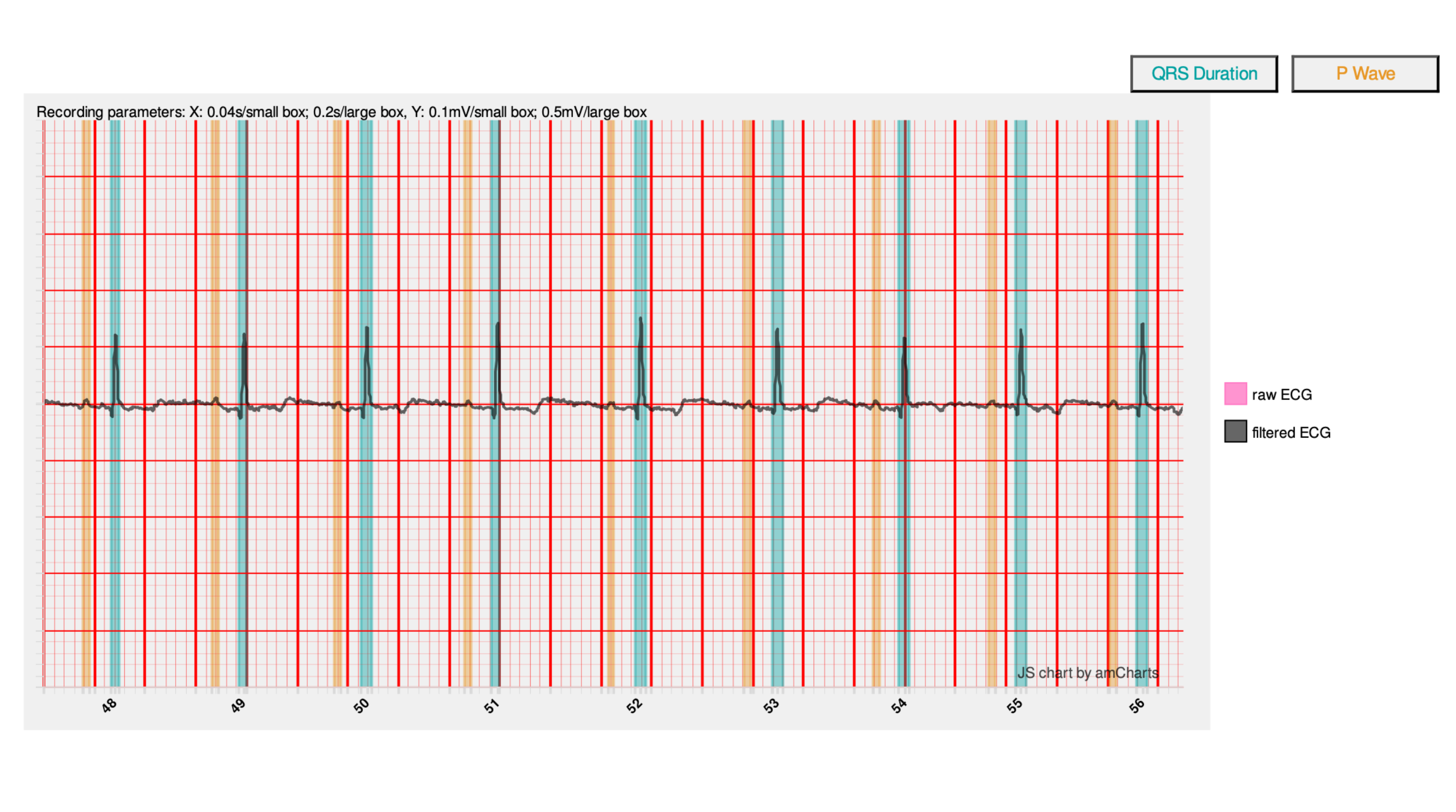
Figure 1. Lead II ECG strips (selected segments). This figure illustrates the irregular RR intervals, preserved P‑QRS‑T sequence, and the prolonged PR and QRS durations detected by CardioBird.
ECG findings and interpretation
The AI‑assisted measurements showed a mean PR interval of 0.104 s, exceeding the feline reference range of 0.05–0.09s and consistent with first‑degree atrioventricular (AV) block. P waves preceded every QRS complex, and there was no evidence of dropped beats, supporting a diagnosis of first‑degree AV block rather than higher‑grade conduction disturbance.
QRS complexes were mildly prolonged, with a mean duration of 0.041 s and prolongation documented in 48 of 63 beats, relative to the normal feline upper limit of ≤0.04 s. Despite this, both P and R wave amplitudes remained within reference limits (P wave average amplitude 0.065 mV, standard <0.2 mV; R wave average amplitude 0.697 mV, standard <0.9 mV), which reduces suspicion of marked chamber enlargement but does not exclude it.
CardioBird flagged sinus bradycardia with irregular rhythm, first‑degree AV block, and QRS prolongation as the key abnormalities. In a cat without overt clinical signs, this combination strongly suggests increased vagal tone, which may be situational, drug‑related, or associated with extracardiac disease affecting intracranial, ocular, respiratory, gastrointestinal, or urogenital systems. However, the QRS prolongation also raises the possibility of ventricular enlargement or bundle branch block, warranting structural evaluation.
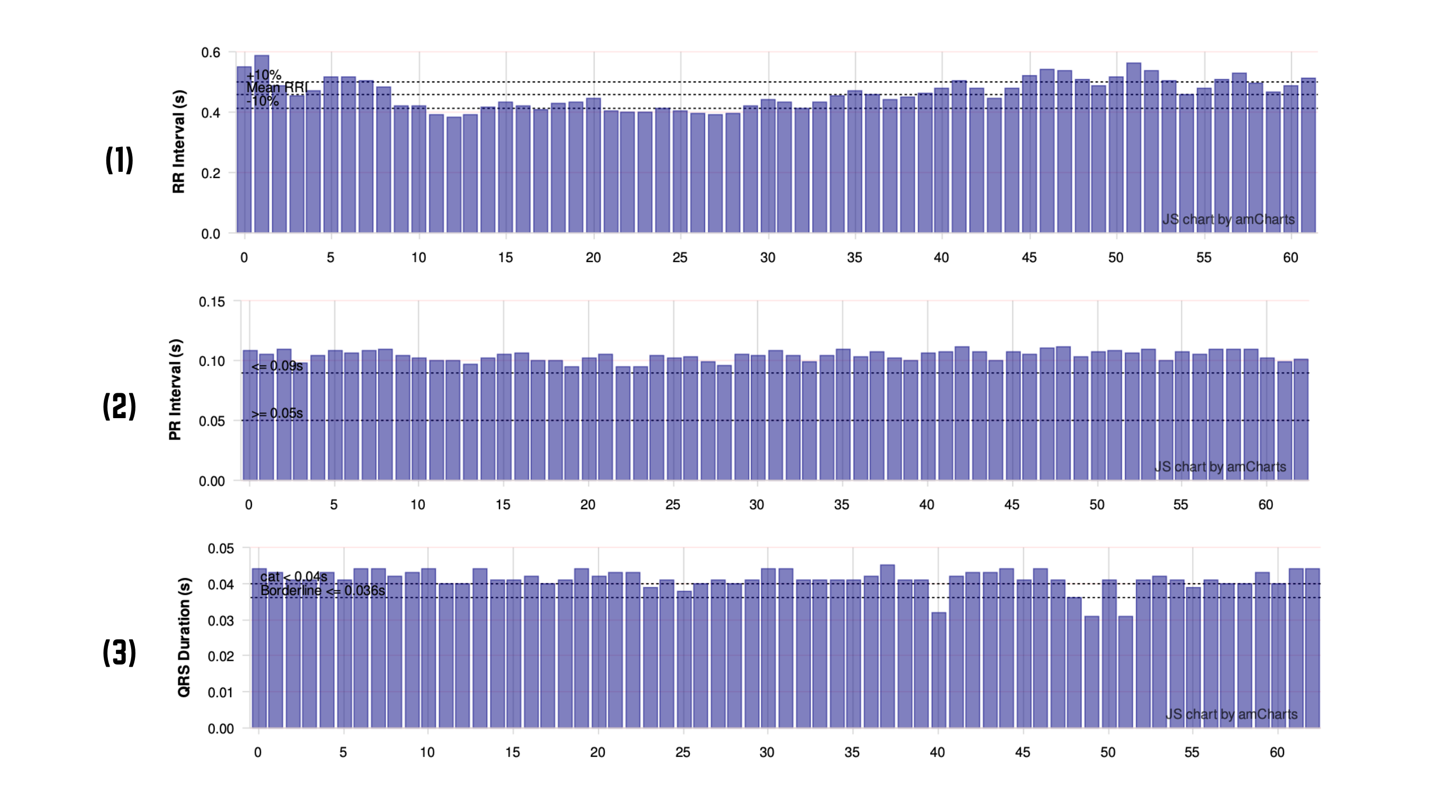
Figure 2. CardioBird interval charts. The RR‑interval plot, and PR/QRS duration charts demonstrate: (1) beat‑to‑beat RR variability; and (2) a shift of PR and (3) QRS values above reference limits in a significant proportion of beats.
Further investigations indicated
In a young adult Maine Coon with conduction abnormalities, the threshold for further work‑up should be low, even in the absence of clinical signs. Maine Coons are a breed in which structural heart disease, including hypertrophic cardiomyopathy, is an important consideration, and electrical changes may precede overt changes on physical examination.
Recommended next steps include:
- Thoracic radiography
To assess cardiac silhouette size and pulmonary vasculature and to look for evidence of cardiomegaly or other thoracic pathology that could explain increased vagal tone (e.g. respiratory disease). - Echocardiography
To define chamber dimensions, wall thickness, systolic function, and outflow tracts, and to identify occult cardiomyopathy or regional conduction‑related abnormalities (e.g. focal wall motion changes). - NT‑proBNP measurement
To add an objective biomarker of myocardial stress, helping to stratify risk and guide the urgency of cardiology referral. - Minimum database and electrolytes
To exclude systemic or metabolic contributors to bradycardia and conduction delay, including electrolyte imbalance and drug effects. - Atropine response test (where appropriate)
To differentiate vagally mediated sinus bradycardia and first‑degree AV block from intrinsic conduction system disease.
These investigations align with the pathways suggested in the CardioBird report and help veterinarians move from pattern recognition on ECG to a complete, clinically integrated diagnosis.
Specialist’s suggested clinical management
For this cat, management should proceed along two parallel tracks: (1) immediate risk assessment and safety, and (2) longer‑term monitoring and decision‑making guided by structural and biomarker findings.
In the short term, if the cat remains asymptomatic with no syncope, weakness, or signs of congestive heart failure, outpatient evaluation is appropriate while avoiding unnecessary negative chronotropic or dromotropic medications. Any sedatives, anaesthetics, or other drugs that may exacerbate bradycardia or AV nodal delay should be reviewed and adjusted where feasible.
Once thoracic imaging, echocardiography, and NT‑proBNP results are available, management diverges:
- If structural heart disease is identified
Treatment should follow current cardiomyopathy guidelines for cats, with ECG findings used as an additional marker of disease severity and a baseline for future comparison. - If no significant structural or systemic disease is found
The first‑degree AV block and mild QRS prolongation can be monitored as likely manifestations of high vagal tone, with repeat ECGs scheduled (e.g. at 6‑month intervals, or sooner if clinical signs develop). Serial ECGs can detect progression to higher‑grade block, worsening QRS prolongation, or evolving arrhythmias.
Owner communication is crucial: explaining that the ECG has uncovered early electrical changes at a stage when the cat feels well underscores the value of routine ECG screening and supports adherence to follow‑up recommendations. For veterinarians, this case exemplifies how AI‑enhanced ECG—integrated into wellness visits—can refine risk stratification, prompt timely imaging, and create a robust baseline for lifelong cardiac care.
Want to see the full analysis for this Maine Coon?
Scan the QR code below, and send us a message to access the Full CardioBird AI‑ECG report and explore the interactive charts.